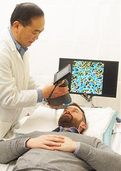
Dermatologists may soon be able to carry out evaluations and make diagnoses quickly and accurately without the need for tissue samples or injectable dyes.
 Wausau, WI (March 27, 2018) – Visually confirming the presence of a disease within a tissue frequently involves a sample of tissue being taken and studied. This technique, however, is invasive, time consuming and requires additional trauma meaning monitoring over time is not ideal. Another option growing in popularity is the study of blood vessels. Research has shown that such vessels change in response to the presence of a disease and each disease type will cause the vessels to change in different ways. The degree of change is also often an indication of how severe the disease is at that time. Currently, these vessels are largely visualized with the use of injected dyes that can be seen through a monitor when light is shone on the body. However, this use of dyes is again not ideal.
In a study published in Lasers in Surgery and Medicine (LSM), the official journal of the American Society for Laser Medicine and Surgery, Inc. (ASLMS), a team of researchers adapted an imaging technique that uses light to visualize the vessels in the skin in a similar way to how ultrasound uses sound waves. This technique, optical coherence tomography angiography (OCTA), is commonly used to visualize the vessels of the eye; however, they developed a specific clinically translatable system to image both the vessels and structure of the skin. For this study, the healthy skin of countless volunteers, numerous common skin conditions, such as moles and skin tags, and a number of diseases, such as psoriasis, graft-versus-host disease (GvHD), and scleroderma were imaged. In each case, it was shown that the vessels of the skin differ depending upon their location on the body and/or the presence of a particular disease. Additionally, they were able to match vessel features with structural features, meaning a visual inspection by a dermatologist can be easily correlated with certain vessel characteristics under the surface of the skin.
The study was conducted by Ruikang K. Wang, PhD; Faezeh Talebi-Liasi, MD; Shaozhen Song, PhD; Yuandong Li, MSc; Jingjiang Xu, PhD; Shaojie Men, PhD; Michi M. Shinohara, MD; Mary E. Flowers, MD; Stephanie J. Lee, MD. Their manuscript titled, “Optical Coherence Tomography Angiography of Normal Skin and Imflammatory Dermatologice Conditions” was selected as Editor’s Choice in the March 2018 issue of LSM.
In all, the OCTA system allows vessels and structural features of the skin to be visualized with a level of clarity that has not been seen before.
“Optical coherence tomography angiography allows us to visualize the microvessels and structural features within the skin tissue beds with a level of clarity that has not been seen before,” said Dr. Wang.
“This technique has the potential to become commonplace in dermatologic clinics and it is hoped that our system will soon lead to the development of an imaging practice that will allow the dermatologists to carry out evaluations and make diagnoses quickly and accurately without the need for tissue samples or injectable dyes.”
Dr. Wang is a full professor with appointments in the Departments of Bioengineering and Ophthalmology at the University of Washington and directs the Biophontonics and Imaging Laboratory. He is an elected fellow of Optical Society of America (OSA), International Society for Optics and Photonics (SPIE) and American Institute for Medical and Biological Engineering (AIMBE). His current research interests include biophotonics and imaging, optical coherence tomography, optical microangiography, photoacoustic imaging and their applications in ophthalmology, dermatology, neuroscience and cancer. He has published more than 350 peer reviewed journal articles in the fields of his interests and holds more than 10 patents related to the OCT/OCTA technology.
Editor’s Choice is an exclusive article published in LSM, the official journal of the ASLMS. View the complete manuscript.
The American Society for Laser Medicine and Surgery, Inc. (ASLMS) is the largest multi-disciplinary professional organization, dedicated to the development and application of lasers and related technology for health care applications. ASLMS promotes excellence in patient care by advancing biomedical application of lasers and other related technologies worldwide. Currently, ASLMS has over 4,000 members, including physicians and surgeons representing more than 51 specialties, physicists involved in product development, biomedical engineers, biologists, nurses, industry representatives and manufacturers. For more information, visit aslms.org.