find a Clinician/Physician

Past Research Grant Recipients

To view past research grant and student research grant recipients, click on the "+" to expand each box. 


  • Past Recipients - 2021

    2021 Research Grant Recipients


    singh-rupesh

    Rupesh Singh, PhD

    Cleveland Clinic Cole Eye Institute

    Supporting ASLMS Member: Shilpi Khetarpal, MD

    “Development of a novel therapeutics for nonresponsive neovascular age-related macular degeneration”


    2021 Student Research Grant Recipients


    dos-anjos-carolina 

    Carolina dos Anjos, MSc, DVM

    Massachusetts General Hospital

    Supporting ASLMS Member: R. Rox Anderson, MD

    “Administration of oxygen microbubbles and antimicrobial blue light for the treatment of urinary tract infections”


    prasad-karthik

    Karthik Prasad, BS

    UCI Health Beckman Laser Institute and Medical Clinic

    Supporting ASLMS Member: Brian J.F. Wong, MD, PhD

    “Multimodal Optical Characterization of Electromechanical Cornea Reshaping”


  • Past Recipients - 2020

    2020 Research Grant Recipients
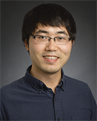
     

    gao-liangLiang Gao, PhD

    The Regents of the University of California, Los Angeles

    Supporting ASLMS Member: Brian J.F. Wong, MD, PhD

    “Label-free Real-time Hyperspectral Autofluorescence Lifetime Imaging for Molecular-guided Surgery of Oral Cancers”
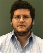

    hajjarian-zeinab

    Leon Leanse, PhD

    Massachusetts General Hospital

    Supporting ASLMS Member: R. Rox Anderson, MD

    “Blue light makes antibiotics great again!”


    2020 Student Research Grant Recipients
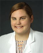
    carney-bonnie

    Bonnie C. Carney

    MedStar Health Research Institute

    Supporting ASLMS Member: Taryn E. Travis, MD, FACS

    “Treatment of hypopigmented hypertophic scar with synthetic alpha melanocyte stimulating hormone (α-MSH) through micro-channel drug delivery”


    kawa-nisrineNisrine Kawa, MD

    Wellman Center for Photomedicine, Massachusetts General Hospital

    Supporting ASLMS Member: R. Rox Anderson, MD

    “Effectiveness of small-scale Z-incisions with photochemical tissue bonding closure in reduction of skin tension"


    li-yanYan Li

    The Regents of the University of California

    Supporting ASLMS Member: Yona Tadir, MD

    "Monitoring and Management of Vaginal Health via Multifunctional OCT/OCTA/OCE Endoscopy"
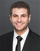

    mehrabi-joseph-nJoseph N. Mehrabi, BS, MS

    Department of Dermatology at University of California, Irvine

    Supporting ASLMS Member: Christopher B. Zachary, MBBS, FRCP

    “Optical coherence tomography guided laser treatment of non-melanoma skin cancers”


  • Past Recipients - 2019

    2019 Research Grant Recipients

    hajjarian-zeinab

    Zeinab Hajjarian, PhD

    Massachusetts General Hospital
    Wellman Center for Photomedicine
    Harvard Medical School

    Supporting ASLMS Member: R. Rox Anderson, MD

    "Identifying an Early Mechanbo-biomarker of Metastasis Risk in Breast Carcinoma Using Laser Speckle Micro-rheology of the Tumor Extra-Cellular Matrix"

    Our goal is to exploit a novel optical approach for micro-mechanical imaging of the breast tumor tissue and for developing an early mechano-biomarker of metastasis risk in patients.

    yin-rong

    Rong Yin, PhD

    University of South Carolina School of Medicine

    Supporting ASLMS Member: J. Stuart Nelson, MD, PhD

    "Targeting Port Wine Stain Blood Vessels by Endothelial Optical Exosomes"

    The purpose of this seed grant aims at engineering novel endothelial exosome-derived optical nanoparticles for treatment of Port wine stain (PWS), which can be ultimately developed as a personalized precision photodynamic therapy (PDT) for congenital vascular malformations (CVM).

    2019 Student Research Grant Recipients

    ahluwalia-jusleen

    Jusleen Ahluwalia, MD

    UC San Diego

    Supporting ASLMS Member: Arisa E. Ortiz, MD

    "Topical Tranexamic Acid Versus Fractional Drug Delivery of Tranexamic Acid for the Treatment of Melasma"

    We hypothesize that the fractional Er:YAG delivery of TA 5% cream would result in a greater decrease in MASI scores when compared with topical TA alone given its multitargeted approach with efficient drug delivery, and anti-tyrosinase activity of TA. Because the cutaneous absorption of the Er:YAG laser by water is more efficient than the CO2 laser, the Er:YAG laser has potential for less thermal damage and thus decreased risk to develop post-inflammatory hypopigmentation. We hypothesize that this combination approach will provide an effective, long-term treatment option for patients suffering from melasma.

    chen-jason

    Jason Chen

    The Regents of the University of California

    Supporting ASLMS Member: Brian J.F. Wong, MD, PhD

    "Quantification of Symptomatic Vitreous Floaters Using Optical Coherence Tomography"

    We first aim to utilize VCSEL as the light source to achieve near full-depth imaging of the vitreous body for the quantification of floater geometry. Secondly, we will address the challenges associated with increasing imaging ranges, namely increased dispersion and limited depth of focus.

    nathan-neera

    Neera Nathan, MD

    Massachusetts General

    Supporting ASLMS Member: Molly Wanner, MD, MBA

    "The clinical, microscopic and molecular effects of ablative fractional CO2 laser-assisted drug delivery of collagenase on striae distensae"

    We hypothesize that ablative fractional CO2 laser-assisted drug delivery of collagenase will improve the appearance of striae distensae more than ablative fractional CO2 laser alone. We propose that the mechanism of clinical improvement will be secondary to potentiated remodeling of dermal collagen, elastin and extracellular matrix components by this combination treatment, as evidenced by microscopic and molecular markers of normalization of collagen, elastin, fibrillin, MMPs and TGF-β.

    quang-tri

    Tri Quang

    The University of Iowa

    Supporting ASLMS Member: Yang Liu, PhD

    "Real-time Tissue Perfusion Assessment Using Fluorescence Imaging Topography Scanning System"

    The objective of this proposal is to develop a clinically useful FITS system for intraoperative blood perfusion and assessment, and to evaluate the feasibility of the developed FITS prototype for real-time visualization of vascular flow in small animal models.

    rosenthal-amanda

    Amanda Rosenthal, MD

    Moy-Fincher-Chipps Facial Plastics & Dermatology

    Supporting ASLMS Member: Ronald Moy, MD

    "Effects of 2,940-nm Fractional Ablative Erbium and Topical DNA Repair Enzymes on p53 Epidermal Expression After 3 months: A Comparative Clinical Trial"

    The objective of this study is to compare the impact of topical DNA repair enzymes, specifically T4 Endonuclease V, and laser resurfacing with the 2,940nm fractional erbium on epidermal p53 expression.

    sawyer-travis

    Travis Sawyer

    University of Arizona

    Supporting ASLMS Member: Jennifer Kehlet Barton, PhD

    "Combined Optical Coherence Tomography and Autofluorescence Imaging for Screening of Early-stage Esophageal Cancer"

    The ultimate outcome of this study is to determine the diagnostic potential of combined OCT-AFI for disease screening. If positive, this could create a new paradigm for esophageal tissue assessment, reducing the number of biopsies necessary to successfully identify a case of early-stage cancer. In addition, given the clinically-translatable design, a positive result will motivate the pursuit of an RO1 grant to conduct a clinical trial.

  • Past Recipients - 2018

    2018 Research Grant Recipients

    Leanse-Leon-G

    Leon G. Leanse, PhD

    Massachusetts General Hospital
    Wellman Center for Photomedicine

    Boston, MA

    Supporting ASLMS Member: R. Rox Anderson, M.D.

    “Synergistic action of antimicrobial blue light and quinine against multidrug-resistant bacterial infections: Efficacy and molecular mechanisms”

    With the rise in antimicrobial resistance, it is essential that we look to other, ‘non-antibiotic’ strategies, e.g. antimicrobial blue light (aBL), as alternative methods to treat infections. aBL therapy is a novel and effective approach for the treatment of infections caused by different pathogens. Although aBL has been shown to be highly effective in inactivating pathogens, varied sensitivities are often observed between microbial species, with some eliciting significantly high relative resistances. Therefore, it is vital that approaches, such as aBL combination therapies, are investigated, so we may improve microbial inactivation while simultaneously providing safer and more timely care to patients.

    2018 Student Research Grant Recipients

    Hosking-Anne-Marie

    Anna-Marie Hosking, MS

    University of California, Irvine – Beckman Laser Institute

    Irvine, CA

    Supporting ASLMS Member: Kristen M. Kelly, MD 

    “Spatial Frequency Domain Imaging for Burn Wound Severity Assessment”

    The proposed study will be the first to describe the use of spatial frequency domain imaging (SFDI) in the in vivo evaluation of burn patients. This study will prospectively evaluate burn patients with SFDI in addition to standard care and evaluation by a burn surgeon.

    Molina-Stephanie

    Stephanie Molina, BS

    Board of Regents, NSHE, obo University of Nevada, Las Vegas

    Las Vegas, NV

    Supporting ASLMS Member: Steen Madsen, PhD

    “Macrophage-Mediated Drug Delivery for the Treatment of Malignant Gliomas by PCI”

    This project proposes the use of macrophage-delivered chemotherapeutics combined with photochemical internalization (PCI) to enhance cell-killing efficacy in a glioma cell monolayer. Translation of macrophage-mediated delivery to clinical application will result in site-specific distribution of large drug concentrations within the tumor microenvironment.

    zuccaro-jennifer

    Jennifer Zuccaro, MSc

    Hospital for Sick Children (SickKids)

    Toronto, ON, Canada

    Supporting ASLMS Member: Joel S. Fish, MD, MSc, FRCS(c

    “A Pilot Study to Treat Minor Burn Scar Contractures Using Laser Therapy"

    The primary objective of this study is to determine is using an ablative carbon dioxide laser (ACL) in surgical mode is effective in improving the active range of motion of a joint affected by a minor burn scar contracture as measured by goniometry in children aged 1 to 18 years.

  • Past Recipients - 2017

    2017 Research Grant Recipients

    Vermandel-Maximilien

    Maximilien Vermandel, PhD

    Inserm

    France

    “INDYGO-Intraoperative PDT for the Treatment of Glioma”

    Our main objective concerning biomarkers within this application is to obtain new data on the effectiveness of the PDT by monitoring MRI.  Additionally, biological samples will be collected during the trial to enable future exploratory studies on the evolution of immune cell subpopulations, the secreted factors and the nanovesicules released into the blood.  All together these data should allow future identification of early biomarkers of treatment response.


    2017 Student Research Grant Recipients

    Ford-Jeremy

    Jeremy Ford, BE

    Vanderbilt University

    Nashville, TN

    “A Custom Probe for Reducing Thermal Load During Infrared Neural Inhibition"

    Our goal is to evaluate whether applying spatially and temporally variant thermal profiles to nerves will increase inhibition efficacy and reduce tissue heat load, thus making Infrared neural inhibition (INI) clinically feasible.

    Mehta-Jason-N

    Jason N. Mehta

    The University of Texas at Austin

    Austin, TX

    “Remote Control System for Fiberoptic Microneedle Device Catheter”

    We are developing and prototyping an “arborizing fiberoptic microneedle device (FMD)’ and a remote control interface that can be operated by a neurosurgeon while the patient is observed by the use of a commercial MRI scanner.

  • Past Recipients - 2016

    2016 Research Grant Recipients

    arany-praveen-r

    Praveen R. Arany, BDS, MDS, MMSc, PhD

    The Research Foundation for The State University of New York on behalf of University at Buffalo

    Albany, NY

    “Functional clinical validation of Photobiomodulation Therapy for Treatment of Traumatic Brain Injury”

    This proposal will attempt to establish therapeutic benefits of low dose clinical biophotonics, termed Photobiomodulation (PBM) therapy, in treating traumatic brain injury (TBI). In this pilot human study, we will rigorously evaluate neurocognitive, functional, and structural deficits present in a group of TBI patients and perform PBM treatments to assess preliminary therapeutic efficacy. 

    Siddhanta-Soumik

    Soumik Siddhanta, PhD

    Johns Hopkins University

    Baltimore, MA

    “Targeted In Vivo Detection of Breast Lesions and Microcalcifications with Raman Spectroscopy

    Our proposed research is focused on integrating vibrational spectroscopy with existing biopsy equipment to create a minimally invasive biopsy guidance system that can definitively inform the radiologist in real-time about the pathologic nature of the tissue cores to be biopsied. We ultimately envision that integrating the rich molecular detail of Raman spectroscopy with spatially offset probes and tomography can offer fully non-invasive chemical characterization and microstructure visualization (“optical biopsy”).


    2016 Student Research Grant Recipients

    Alhallak-Kinan

    Kinan Alhallak

    University of Arkansas

    Fayetteville, AR

    “Diffuse Optical Spectroscopy to Distinguish Between Metastatic and Non-Metastatic Breast Cancers”

    Our goal is to determine the differences in oxygenation and glycolytic demand of primary breast tumors with varying metastatic potential. Specifically, we hypothesize that highly metastatic tumors will present with significantly elevated glycolytic demand coupled with high levels of oxygenation compared with non-metastatic tumors.

    Pal-Rahul

    Rahul Pal, MS

    University of Texas Medical Branch at Galveston

    Galveston, TX

    “Multispectral Characterization of Wide-Field Autofluorescence for Multimodal Imaging in Detection of Oral Pre-Neoplasia in a Hamster Oral Cancer Model”

    This project explores the innovative and previously unexplored concept of multispectral imaging to identify high-risk pre-cancerous tissue outside of clinically visible tumor margin.   Imaging will be performed using a novel liquid crystal tunable filter for multispectral image acquisition and sequential illumination with multiple excitation wavelengths where characteristic contribution from different tissue components will be visible.

    Yao-William

    William Yao, PhD

    The Regents of the University of California, Los Angeles

    Lost Angeles, CA

    “Establishing a Therapeutic Window for Laser Generated Shockwave Treatment for Bacterial Biofilms”

    We aim to demonstrate the preclinical efficacy of laser generated shockwaves (LGS) as an adjunct therapy in the management of cutaneous biofilms, fueling a paradigm shift towards safe, less time intensive therapy of infected wounds. This technology has potential application to a wide range of cutaneous chronic infections, where the current therapy requires repeated painful debridement and prolong wound care.

  • Past Recipients - 2015

    2015 Research Grant Recipients

    Li-Ming

    Ming Li, PhD

    Postdoctoral Fellow

    Mechanical Engineering
    Johns Hopkins University

    Baltimore, MD

    “A Photonic Tool for Targeted Theranostics of Prostate Cancer Using a Urea-Based Small-Molecule Inhibitor”

    Our goal here is to design, develop and validate this novel theranostic nanoplex in cellular systems and in mice models using a high-speed Raman microscopy system. In particular, we first seek to architect and synthesize a surface-enhanced Raman spectroscopy (SERS) probe based on gold nanostar cores and conjugate with prostate-specific membrane antigen (PSMA) targeting moiety.  Subsequent to validation in PSMA over-expressing cell lines, we will link the beta-particle radiotherapeutic moiety and test the combined nanoplex in mouse xenograft models. Our findings here would also provide pharmacokinetics, toxicity and immunogenicity insights of the theranostic nanoplex. Taking the complementary expertise in our team, our present efforts will provide a reliable tool for prostate cancer care and management via SERS image-guided surgery and cancer-selective treatment.

    Mallidi-Srivalleesha

    Srivalleesha Mallidi, PhD

    Research Fellow

    Harvard Medical School
    Wellman Center for Photomedicine
    Massachusetts General Hospital

    Boston, MA

    “Treatment of Residual GBM With Image-Guided PDT and Multi-Agent Nanoconstructs”

    Scientifically this proposal provides a nanoplatform for the selective co-delivery of multiple independently compartmentalized agents that will allow for rapid evaluation of a broad array of therapeutics and mechanisms through photoacoustic imaging techniques. Clinically, these findings will enable patient-specific, image-guided drug delivery and will advance options for the treatment of residual glioblastoma multiforme (GBM), the major cause of recurrent disease and BGMB-related mortality.


    2015 Student Research Grant Recipients

    Ganguly-Mohit

    Mohit Ganguly

    Graduate Student Researcher

    Biophotonics Center
    Biomedical Engineering Vanderbilt University

    Nashville, TN

    “Photonic Analgesia by Inhibition of Nerves (PAIN)”

    We plan to develop optical nerve inhibition as a means of precise (i.e. blocking only the necessary portion of a nerve) and non-pharmacological nerve inhibition for modulating neural activity, thereby inhibiting both acute and chronic pain.

    photonotavailabe_000

    Bryan Lemieux

    Student Researcher, MS4

    University of California Irvine School of Medicine
    Beckman Laser Institute

    Irvine, CA

    “In Vivo Measurement of Ciliary Beat Frequency Using Optical Doppler Tomography”

    The objectives of this study are to develop an OCT system and probe can be used to measure ciliary beat frequency (CBF) using Doppler technology; use this system to obtain high frame rate images in vivo in the rabbit upper airway; and develop a program in MatLab to extract CBF data from these high frame rate images and map CBF in the upper airway.  This in vivo model will serve as a first step toward translating this technology to clinical use.

    Mac-Jenny

    Jenny Mac

    Graduate Student Researcher

    The Regents of the University of California, on behalf of Riverside Campus

    Riverside, CA

    “Erythrocyte-Derived Nanoparticles for Targeted Near-Infrared Imaging and Laser-Mediated Photothermal Destruction of Cancer Cells”

    As a first step to determine the efficacy of NETs for photothermal destruction of cancer cells, here I propose studies directed at targeted destruction of cancer cells using NETs in-vitro.  The human epidermal growth factor receptor-2 (HER2) is overexpressed in a range of tumor types including breast, ovarian, and pancreatic cancer.  In particular, I will be functionalizing NETs with anti-HER2/neu to target ovarian cancer.  Successful outcomes of these experiments will provide the proof-of-concept data to further pursue NETs for imaging and photothermal destruction of cancer in animal models, and ultimately in patients.

    Wang-Yucheng

    Yucheng Wang

    Graduate Research Assistant

    Massachusetts General Hospital

    Boston, MA

    “Antimicrobial Blue Light Inactivation of Biofilms: In Vitro and In Vivo Studies”

    In this application, we will first carry out in vitro studies on the efficacy of antimicrobial blue light inactivation of biofilms formed by a wide range of clinically important bacterial species, with multiple MDR strains of each species.  Then, by using a mouse model of urinary tract infection (UTI), we will investigate the efficacy of antimicrobial blue light inactivation of biofilms in vivo.

  • Past Recipients - 2014

    2014 Research Grant Recipients

    Ortiz_Arisa_jpg

    Arisa Ortiz, M.D.

    University of California

    San Diego, CA

    Supporting ASLMS Member: R. Rox Anderson, M.D.

    “Long-Pulsed 1064nm Nd:YAG Laser Treatment of Basal Cell Carcinoma”

    Basal cell carcinoma (BCC) is the most common skin cancer worldwide. Although this tumor is rarely life threatening, it is locally destructive and can cause significant cosmetic and functional morbidity. Standard therapeutic modalities for BCC often result in disfigurement from surgical treatments and recurrences with topical therapies. Thus, there is a need for alternative non-surgical options that are effective, efficient, and have a low risk of side effects. This has led to the emergence of laser investigations for the treatment of BCC due to the ease of treatment, lack of significant downtime, decreased risk of complications, and absence of a surgical scar. The primary objective of this study is to evaluate the safety, clinical and histological efficacy of high-fluence 1064 nm Nd:YAG laser therapy for the treatment of subjects with BCC on the trunk and extremities that do not meet the criteria for Mohs Micrographic Surgery.


    White_Sean_tif

    Sean White, Ph.D.

    University of California

    Irvine, CA

    Supporting ASLMS Member:Bernard Choi, Ph.D.

    "Development of Clinic-Friendly Perfusion Measurement System to Aid in Light Dosimetry During Novel Port Wine Stain Treatment"

    Port-wine stain (PWS) birthmarks affect approximately ~400,000 individuals annually and are characterized by potentially deleterious psychosocial and physical complications. The current standard of care for these lesions, photocoagulation using a pulsed dye laser (PDL), achieves unacceptable PWS lightening in >85% of subjects even after multiple treatments. Limited treatment efficacy is due in part to the fact that (1) PDL irradiation does not effectively photocoagulate blood vessels with diameters <20 μm; (2) epidermal melanin reduces the light dose reaching the target vessels; and (3) light dosimetry is often based solely on clinical experience, which can lead to inadequate or excessive energy deposition during treatment. To address the first and second shortcomings, we are currently conducting a clinical trial to investigate the efficacy of a novel dual-modality treatment for PWS. By utilizing photodynamic therapy (PDT) in addition to PDL irradiation, melanin’s absorption of incident light can be mitigated with proper wavelength choice, and blood vessels of all sizes can be treated with a smaller light dosage than would be required for PDL monotherapy. As such, the dual-modality treatment may increase PWS blanching while minimizing potential side-effects such as scarring. To address the third shortcoming, we propose to expand upon previous work by building a real-time perfusion measurement system for use during PWS phototherapy. This system will be clinician-operable and offer real-time dosimetry feedback during treatment. To accomplish this goal, we will first construct a cutaneous perfusion imaging system based upon the principal of laser speckle imaging. This system will benefit from our group’s extensive experience in creating customized laser speckle imaging system hardware and software, particularly exemplified by two previous systems created to monitor PWS perfusion during PDL treatment. We will subsequently use this perfusion measurement system to quantify the reduction in PWS perfusion during dual-modality treatment, as well as during PDT and PDL monotherapy. Treatment efficacy will be quantified as the degree of PWS blanching (measured using calibrated color photography and colorimetry), and the correlation between treatment efficacy and the decrease in perfusion will be computed for each treatment type. With this data, clinicians can then use the perfusion measurement system to quantitatively determine a treatment’s endpoint by targeting the drop in PWS perfusion that corresponds to the desired degree of PWS blanching. Clinicians will thus have the ability to quantitatively determine when treatment should cease, reducing the risk of under- or over-treating PWS lesions, therefore ameliorating the efficacy of each treatment session on an individualized basis.


    2014 Student Research Grant Recipients

    FarraroRyan_tif_000

    Ryan T. Farraro

    University of California

    Irvine, CA

    Supporting ASLMS Member

    Bernard Choi, Ph.D.

    “Handheld Laser Speckle Imaging System for Neonatal Care”

    The longer a neonate stays in a hospital, the greater their risk of developing an infection; therefore, to address the urgent treatment of neonatal diseases, there is a need for more early diagnostic devices for neonates. Laser speckle imaging is an ideal solution to this problem as it allows for real-time, non-invasive assessment of abnormal microvascular flow— a vital parameter that has been linked to several neonatal diseases. While laser speckle imaging has been used in a clinical setting, it has not been used as a point-of-care device. The goal of this proposed research is to assess whether or not a handheld laser speckle imaging device can be used in a clinical setting to take blood flow measurements of neonates. In order to accomplish this, the device must be characterized to understand the limitations of a handheld device. Characterization will be performed in three stages: 1) understanding the effect of movement on the speckle contrast images, 2) quantitatively measuring sensitivity to flow, and 3) performing a preliminary study within the hospital. Once the device has been characterized, a future study can be undertaken using the device to study neonatal disease states.

    Pence_Isaac_jpg

    Isaac Pence

    Vanderbilt University

    Nashville, TN

    Supporting ASLMS Member

    E. Duco Jansen, Ph.D.

    “In Vivo Raman Spectroscopic Characterization of Active Inflammatory Bowel Disease”

    The development of Raman spectroscopy as a real-time, optical diagnostic tool for integration with conventional video endoscopy to differentiate inflammatory bowel disease (IBD) classes will be researched. Initial research for this project included establishing spectral biomarkers in ex vivo colon biopsy samples to differentiate ulcerative colitis and Crohn’s disease from normal controls. Furthermore, we have developed an optimized, endoscope-compatible Raman fiber probe design to enable in vivo data collection. The proposed research project, to characterize the in vivo disease and normal signals of the colon for IBD, will guide ongoing research and diagnosis of this poorly understood disease. If successful in establishing Raman spectral discrimination of IBD, this technology can be used for real-time in vivo diagnosis and follow-up of chronic IBD patients, providing biochemical information as a diagnostic “gold standard” and improve upon the current inexact clinical, endoscopic, and histological evaluations. The proposed research will be performed concurrently with additional in vivo human measurement studies to compare the relative influence of disease presentation in location activity and other patient variables to establish the accuracy, sensitivity, and specificity of the Raman spectroscopic approach to predicting IBD class.

    regan_caitlin_JPG_001

    Caitlin Regan

    University of California

    Irvine, CA

    Supporting ASLMS Member

    Bernard Choi, Ph.D.


    “Laser Speckle Imaging to Determine Tooth Viability”
    The proposed research project is to design, build, and test a handheld LSI device for measuring pulsatile flow in the tooth to determine viability. All research will be carried out at Beckman Laser Institute at the University of California in Irvine. Initial device design and building, as well as all data processing will be carried out in the lab of Dr. Bernard Choi, who specializes in LSI. Testing the device in human subjects will be performed under the direction of Dr. Petra Wilder-Smith, who already has IRB approval for this study, along with endodontist Dr. Jan O’Dell.

    Tang_Jack_jpeg

    Jack Tang
    University of California

    Riverside, CA

    Supporting ASLMS Member

    Bahman Anvari, Ph.D.

    “Optimization of Fabrication Methods for Maximizing the Fluorescence Emission of Optical Nano-Vectors Derived from Erythrocytes”
    Completion of the proposed research will yield fabrication procedures for constructing near-infrared erythrocyte-mimicking transducers (NETs) with optimized emission intensity and fluorescence quantum yield. We plan to inject fluorescence-optimized NETs into mice to assess the efficacy of vascular imaging over extended circulation times in collaboration with Dr. J. Stuart Nelson’s group at the Beckman Laser Institute. We will also functionalize the surface of NETs using PEG-linkers and antibodies. This should add specific cell-targeting capabilities to the nanovectors. Continued researc

  • Past Recipients - 2013

    2013 Research Grant Recipients

    tan.wenbin-photocopy_000

    Wenbin Tan, Ph.D.

    University of California

    Irvine, CA

    Supporting ASLMS Member: J. Stuart Nelson, M.D., Ph.D.

    “Topical Axitinib Combined with Rapamycin to Inhibit the Regrowth of Port Wine Stain Blood Vessels Post Pulsed Dye Laser Treatment ”

    The pulsed dye laser (PDL) is the current treatment of choice for Port Wine Stain (PWS), but the therapeutic outcome is inadequate due to the reformation and reperfusion of PWS blood vessels. We herein propose that combination of two anti-angiogenesis agents, rapamycin (RPM) and axitinib, should inhibit multiple angiogenesis pathways, including blockage of phosphorylation of VEGFRs and PDGFR and phosphoinositide 3-kinase / AKT / the mammalian target of rapamycin (PI3K/AKT/mTOR), thus leading to an effective inhibition of regrowth of blood vessels post PDL.

    Wangtianyi

    Tianyi Wang, Ph.D.

    University of Texas

    Austin, TX

    Supporting ASLMS Member: Thomas E. Milner, Ph.D.

    “Catheter-Based Combined OCT and Two-Photon Luminescence Imaging System for Detection of Thincap Fibroatheroma ”

    Atherosclerosis and plaque rupture leading to myocardial infarction, stroke and progression of peripheral artery disease remain the leading cause of death worldwide. The proposed project is to construct a catheter-based combined OCT-TPL imaging system to simultaneously detect structure and composition of thin-cap fibroatheromas. The catheter-based OCT-TPL system incorporates spectral-domain OCT (1310 nm), TPL imaging (at 760-1040 nm excitation) and a catheter that combine both. A dispersion compressor is utilized to provide transform-limited pulses. A photonic crystal fiber (PCF) is used to transmit single-mode OCT and TPL light simultaneously to/from the sample. TPL emission signals are directed into photomultiplier tubes (PMT1: 640-720nm; PMT2: <570nm) to distinguish macrophages (loaded with nanoparticles) and other plaque components (e.g., elastin fibers and lipid deposits). Successful completion of the proposed study will establish a novel imaging approach to simultaneously detect plaque structure and composition, and will motivate other application possibilities such as tumor-associated macrophage detection in

    external and internal cancers (e.g., cutaneous melanoma, breast, prostate, ovarian, cervical

    and colon cancers).


    2013 Student Research Grant Recipients

    damestani2

    Yasaman Damestani, B.S.

    University of California

    Riverside, CA

    Supporting ASLMS Member

    Guillermo Aguilar, Ph.D.

    “Transparent Cranial Implant for Laser Based Therapy and Imaging of Brain ”

    The long-range goal of the Windows to the Brain (WttB) is to improve patient care by providing a technique for delivery and/or collection of light from the brain, on demand, over large areas, and on a chronically-recurring basis without the need for repeated craniotomies. WttB holds the transformative potential for enhancing the light based diagnosis and treatment of a wide variety of brain pathologies including cerebral edema, traumatic brain injury, stroke, glioma, and neurodegenerative diseases. The proposed study seeks to have a direct and timely implication for improving patient care through replacement of the skull in regions of interest with an optically-transparent yttria-stabilized zirconia (YSZ) cranial implant. Waveguide-like structures (WG) will be written within the implants using an ultra-low fluence femtosecond laser.

    Wes.jpg

    Wesley J. Moy

    University of California

    Irvine, CA

    Supporting ASLMS Member

    Bernard Choi, Ph.D.

    “Depth of Vascular Shutdown Achieved with Photodynamic Therapy”

    The objective of the proposed project is to determine how treatment parameters affect the maximum depth of blood vessel injury and shutdown of blood flow, within a preclinical rat model. We currently are evaluating the effectiveness of a novel light-based treatment, known as photodynamic therapy (PDT), designed to induce selective shutdown of blood vessels. We expect that PDT will improve upon the current treatment approaches currently used by clinicians
    to remove vascular cutaneous birthmarks from patients.

    walsh.alexandra2

    Alexandra Jule Walsh, B.E., M.S.

    Vanderbilt University

    Nashville, TN

    Supporting ASLMS Member

    E. Duco Jansen, Ph.D.

    “Optical Metabolic Imaging of Organoid Cultures Derived from Frozen Tissues to Predict Drug Response ”

    Targeted cancer drugs shrink tumors by modulating cellular protein expression and signaling. However, many patients do not initially respond to these therapies. Clinically, treatment response is measured by tumor size reduction, and ineffective therapies are not identified until weeks after treatment. Sub-populations of cells can resist therapy and lead to relapse later in life. Treatment regimens for unresponsive tumors include more aggressive surgical intervention and experimental therapeutics, such as PI3K and ErbB3 inhibitors. The OMI-organoid system utilizes optical metabolic imaging to interrogate the metabolic shifts induced by drug treatment in primary tumor derived organoid cultures. This technology has the potential to revolutionize patient care by predicting therapeutic response to multiple drugs and drug combinations, rather than relying on retrospective analysis of the administered drug. Organoids derived from frozen tissues would greatly accelerate development of this technology and allow translation to situations where fresh tissues are not feasible.

    welge2

    Weston A. Welge, B.A., B.S., M.S.

    University of Arizona

    Tucson, AZ

    Supporting ASLMS Member

    Jennifer K. Barton, Ph.D.

    “Mild Temperature Hyperthermia Inducing Doppler Optical Coherence Tomography Endoscope for Early Detection of Colorectal Cancer in a Mouse Model ”

    The MTH-inducing Doppler OCT system may allow for earlier colon cancer detection than other perfusion-detecting systems (ultrasound, perfusion CT) due to its high spatial resolution. OCT is also an effective tool for detecting morphological changes in the early stages of colon tumor growth. The proposed Doppler OCT system is capable of imaging the entire colon surface, which is not possible in standard colonoscopies that rely on analyzing biopsied tissues that represent only a small percentage of the colon surface area. Also, as measurements of perfusion are proportional to oxygen uptake in tumors, this system could also be used to monitor radiation and chemotherapy progress.

  • Past Recipients - 2012

    2012 Research Grant Recipients

    Scarcelli.Giuliano

    Giuliano Scarcelli, Ph.D.

    Massachusetts General Hospital/Wellman Center for Photomedicine

    Cambridge, MA

    Supporting ASLMS Member: R. Rox Anderson, M.D.

    “A Pilot Study to Identify “At-Risk” Subjects for Post-LASIK Ectasia Using Brillouin Microscopy”

    The biomechanical properties of the cornea are essential for its function. Corneal collagen fibers provide the mechanical strength to withstand the intraocular pressure; if corneal tissue becomes abnormally weak, corneal ectasia (i.e. thinning and bulging) ensues, causing severe vision degradation. Abnormal weakening of the cornea is a dreaded complication of LASIK surgery. When clinical symptoms manifest, corneal ectasia is often at an advanced stage that leads to corneal transplant. If inherent corneal weakness were detected early, at-risk subjects could be consulted to avoid LASIK surgery; however, current clinical instruments only provide morphological information. This proposal addresses this need through Brillouin microscopy, a novel imaging technology that maps the elastic modulus of material without contact and with 3D resolution. The central hypothesis of the proposal is that low Brillouin elastic modulus is a predictor of post-LASIK ectasia. A pilot clinical study will be performed to compare normal, post-LASIK normal and post-LASIK ectasia corneas in vivo. Introducing elasticity-based risk factors is likely to change the current paradigm for LASIK pre-operative screenings and enable proper identification of at-risk candidates.

    tan.wenbin-photocopy_000

    Wenbin Tan, Ph.D.

    University of California/Beckman Laser Institute

    Irvine, CA

    Supporting ASLMS Member: J. Stuart Nelson, M.D., Ph.D.

    “Molecular Mechanism of Photochemotherapy on Port Wine Stain Disease”

    The proposed research will focus on targeting the clinical barrier that appears to diminish PWS laser therapeutic outcome, namely reformation and reperfusion of PWS blood vessels after PDL treatment. A series of pharmacological, molecular and biochemical approaches are proposed herein to support this novel approach for PWS treatment: (1) the safety, efficacy and pharmacokinetics of newly developed topical formulations of RPM and (2) the time course of RPM-mediated inhibition on angiogenesis pathways induced by PDL. We expect the proposed studies will advance our understanding of molecular pathophysiology of PWS and improve response to PDL treatment.


    2012 Student Research Grant Recipients

    Bahmani.Baharak_000

    Baharak Bahmani, B.S., M.S.

    University of California

    Riverside, CA

    Supporting ASLMS Member

    Jennifer K. Barton, Ph.D.

    “Laser-Mediated Photothermal Destruction of Ovarian Cancer Cells using ICG-loaded Nanocapsules Functionalized with Anti-HER2”

    As a first step to determine the efficacy of ICG-NCs for photothermal destruction of peritoneal ovarian cancer cells, the project is aimed at functionalizing ICG-NCs to target and thermally destroy cancer cells in-vitro. Specifically, due to over-expression of Human Epidermal Growth Factor Receptor 2 (HER-2), ICG-NCs will be functionalized with anti-HER2. Successful outcomes of these experiments will provide the proof-of-concept data to further pursue ICG-NCs for imaging and photothermal destruction of peritoneal ovarian cancer in animal models, and ultimately in patients.

    Tim_Baran

    Timothy M. Baran, B.S.

    University of Rochester – Institute of Optics

    Rochester, NY

    Supporting ASLMS Member

    Thomas H. Foster, Ph.D.

    “Determination of Doxorubicin Concentration in TACE Treatment of Primary Liver Cancers by Fluorescence Spectroscopy”

    Intra-tumor drug concentration can have a large effect on the outcome of the therapy. In previous studies, doxorubicin concentration in systemic circulation was measured. However, the concentration in the tumor was not measured. We propose to study this intra-tumor drug concentration directly by use of fluorescence spectroscopy, since doxorubicin is fluorescent.

    Richa

    Richa Mittal, B.E., M.S.

    University of California

    Irvine, CA

    Supporting ASLMS Member

    Petra Wilder-Smith, D.D.S., Ph.D.

    “A minimally-Invasive Approach to the Challenge of Oral Neoplasia”

    We hypothesize that the photosensitizer hexyl-aminolevulinic (HAL or HexvixR) – a lipophilic ester derivate of 5-Aminolevulinic acid (ALA) - can be used for photodetection and photodestruction of oral dysplasia.

    ASLMSPhoto_ScottThompson

    Scott M. Thompson, B.A.

    Mayo Clinic

    Rochester, MN

    Supporting ASLMS Member

    Roger J. McNichols, Ph.D.

    “Monitoring of Laser Induced Apoptosis with 3D Molecular Bioluminescence Imaging in a Rat Model of Hepatocellular Carcinoma: Pilot Study”

    Our specific aim is to determine the feasibility of non-invasive measurement of apoptotic cell death with combined 3D molecular bioluminescence imaging and magnetic resonance imaging following US-guided laser ablation in a rat model of hepatocellular carcinoma.

  • Past Recipients - 2011

    2011 Research Grant Recipients

    Byrnesmodifiedphoto

    Kimberly R. Byrnes, Ph.D.

    Uniformed Services University

    Bethesda, MD

    Supporting ASLMS Member

    Raymond J. Lanzafame , M.D., M.B.A.

    Modulation of Microglial Activation with Laser Therapy”

    The biological effects of laser therapy (LT), also known as low level light therapy, are currently theorized to follow the Arndt-Schulz law, in which low to moderate energy densities induce activity while higher doses result in inhibition. However, in microglia, there is a range of possible responses, including activation into either a pro-inflammatory (M1) or an anti-inflammatory (M2) state, rather than simple activation and inhibition, excluding them from the Arndt-Schulz law. We therefore aim to show that LT at 810 nm has a differential effect on microglial activation state, depending on energy density. Specifically, we hypothesize that activation of microglia at a low power density will induce an M1 microglial phenotype, while a higher power density will induce an M2 phenotype. These studies will determine if LT can non-invasively modulate microglial phenotype after injury, which would have significant therapeutic potential in the traumatic brain and spinal cord injury fields.

    mathewsphotoforweb

    Marlon S. Mathews, M.D.

    University of California

    Irvine, CA

    Supporting ASLMS Member

    Steen Madsen, Ph.D.

    "Photochemical Internalization (PCI) Enhanced Nonviral Transfection of Tumor Suppressor Genes; a Potential Treatment Modality for Glioma"

    The object of the proposed research is the evaluation and optimization of photochemical internalization (PCI) as a site specific method to increase transfection rates of two tumor suppressor genes, PAX6 and PTEN.


    2011 Student Research Grant Recipients


    photonotavailabe_000

    Adam Boretsky

    University of Texas Medical Branch at Galveston

    Galveston, TX

    Supporting ASLMS Member

    Shelley Sekula-Gibbs, M.D.

    “Adaptive Optics Retinal Imaging of Dry Age-Related Macular Degeneration: Characterization of Cone Photoreceptor Loss and Declining Visual Acuity Associated with Progressive Geographic Atrophy”

    The primary aim of the study described in the current application is to utilize high resolution Adaptive Optics retinal imaging to non-invasively quantify the reduction in macular cone photoreceptor density in patients suffering from dry AMD.

    MdShahidulIslamForWeb

    Md. Shahidul Islam

    University of California

    Riverside, CA

    Supporting ASLMS Member

    R. Rox Anderson, M.D.

    “Development of a Laser Based Optical Electrode for Real Time Non-Contact Monitoring of Nerve Activity”

    The research proposed in this application represents the next step in our careful evolution of this potentially revolutionary technique towards developing an optical electrode for imaging and recording nerve activity in a clinical setting.

    photoforwebalexanderlin

    Alexander J. Lin

    The Beckman Laser Institute / University of California

    Irvine, CA

    Supporting ASLMS Member

    Bernard Choi, Ph.D.

    “Characterizing Optical Signature of Neuron Death in vivo for Alzheimer’s Disease Monitoring”

    The goal of this proposal is to measure and quantify brain changes during Alzheimer’s disease (AD) pathogenesis in a unique transgenic mouse model of inducible cell death.

    nguyenpictureforweb_001

    Tony Nguyen

    The Beckman Laser Institute / University of California

    Irvine, CA

    Supporting ASLMS Member

    Brian J.F. Wong, M.D., Ph.D.

    “Monitoring of Bovine Tendon Structural Changes During Electromechanical Reshaping using Optical Coherence Tomography”

    The objective of this study is to 1) investigate the biomechanical changes (Young’s modulus/ stiffness and changes in strain) of bovine tendon following EMR using flat plate surface electrodes, 2) optimize the process of EMR using needle electrodes to reduce tissue damage and 3) monitor and examine structural changes in the tissue throughout the duration of EMR through optical coherence tomography.

  • Past Recipients - 2010

    2010 Research Grant Recipients

    photonotavailabe_000

    Eduardo H. Moriyama, BSc, MSc, Ph.D.

    Ontario Cancer Institute

    Toronto, Canada

    Supporting ASLMS Member

    James E. Boggan, M.D.

    ~ Recipient of ASLMS Funding in 2009 and 2010 ~

    “Novel Optical Probes for Image-Guided Tumor Resection and Photodynamic Therapy Based on Glucose Transporters”

    Tumors often present a shift in metabolism to a less efficient glycolysis. This has been employed diagnostically, using Fluorodeoxy-D-Glucose Positron Emission Tomography (FDG-PET). We propose to synthesize fluorescently-labeled deoxyglucose analogues (F*-DG), whose potential usefulness is 2-fold. Firstly, they could provide high contrast for fluorescence imaging, in particular to improve fluorescence image guided resection (FGR) of tumors. FGR effectively ‘extends the surgeon’s vision’, so that small amounts of residual tumor that are not normally visible can be detected and removed. We will develop this initially for brain tumors, building on previous work, but the concept is widely applicable to any solid tumor that invades the normal host tissue, such as oral tumors that will be a second target. Secondly, we will test the performance of F*-DG probes as photodynamic sensitizers to improve the effectiveness and selectivity of PDT in these tumors.

    Martin_forweb2

    Martin Purschke, Ph.D.

    Massachusetts General Hospital/Wellman Center for Photomedicine

    Boston, MA

    Supporting ASLMS Member

    R. Rox Anderson, M.D.

    We would like investigate the ATBE with this grant proposal and divide the proposal in two parts: The first part will focus on the mechanism and selectivity of the ATBE by confirming our preliminary data and hypothesis. The second part will focus on in vivo the laser induced ATBE in 3D or skin models to prove the relevance of the ATBE under more complex in vivo conditions (presents of ROS, scavengers and other biomolecules). With the knowledge of the research funded by this grant, we believe it will help in the design of a new laser application for cancer therapy. Laser treatment could be applied, combined or adjuvant to conventional cancer therapies to increase the overall tumor regression. Furthermore, the obtained data may also have profound impact on the treatment paradigms for laser assisted cancer ablation, e.g. laser assisted thermal therapy.


    2010 Student Research Grant Recipients

    dbennettpictureforweb

    David Bennett, BS, MS

    University of California

    Center for Advanced Surgical and Interventional Technology (CASIT)

    Los Angeles, CA

    Supporting ASLMS Member

    Warren S. Grundfest, M.D.


    “Improvement of the Ablation Accuracy of LASIK Using Continuous Terahertz Hydration Monitoring”

    The goal of this research is to demonstrate that hydration monitoring by THz imaging can be used to provide corrective feedback to a clinical excimer laser system in order to increase the ablation accuracy and predictability.


    chenphoto2forweb

    Tina S. Chen, M.D.

    University of California

    Irvine, CA

    Supporting ASLMS Member

    Christopher B. Zachary, M.B.B.S., F.R.C.P.


    “Evaluation of Angiogenesis associated with Laser Treatment of Cutaneous Vascular Lesions”

    The specific objective of this project is to determine the presence of specific angiogenesis mediators in cutaneous vascular lesions before and 4 days after laser treatment. The long term goal is to inhibit production of these growth factors in order to enhance treatment effects. This project has the potential to significantly impact light-based therapeutics.


    schoenly_joshpictureforweb

    Joshua E. Schoenly, BSc

    University of Rochester

    Rochester, NY

    Supporting ASLMS Member

    Raymond J. Lanzafame, M.D., M.B.A.


    “Study of the Selective Ablating Mechanism for Dental Calculus in the Near-Ultraviolet”

    The goal of the proposed research is to measure the selective removal rate (depth of tissue removed per laser pulse) of supra- and sub- gingival calculus in vitro at various laser fluencies (J/cm2).


    EdWuPictureforweb

    Edward C. Kuan, MD, MBA

    University of California

    Irvine, CA

    Supporting ASLMS Member

    Brian J.F. Wong, M.D., Ph.D.


    “Laser Auricular Cartilage Reshaping with Carbon Dioxide Spray Cooling”

    Hypotheses/objectives are to identify optimal parameters to perform LCR with CO2 in ex vivo rabbit auricular tissue; to determine the effects of laser dosimetry and CO2 cooling parameters on: 1) the spatial and temporal evolution of surface temperature; 2) cartilage viability and mechanical behavior; and 3) cutaneous and cartilaginous thermal injury; and to identify and select candidate laser dosimetry and CO2 cooling parameters for auricular reshaping in vivo and evaluate its long-term effects on cartilage tissue.

  • Past Recipients - 2009

    2009 Research Grant Recipients

    CGomerphoto2_000

    Charles J. Gomer, Ph.D.

    Children’s Hospital

    Los Angeles, CA

    Supporting ASLMS Member

    J. Stuart Nelson, M.D., Ph.D.

    “Circulating Endothelial Cells and Circulating Endothelial Progenitors as Biomarkers of Photodynamic Therapy Response”

    The specific objectives of our proposal are to determine if CEC and CEP levels can provide a rapid, reproducible, non-invasive, and clinically relevant prognostic assessment of PDT effectiveness and if targeting CEPs can enhance PDT efficacy. We hypothesize that CEC and CEP measurements will be effective in predicting the efficiency of PDT treatments. CEC levels will serve as a marker of PDT induced vascular damage while CEP levels will serve as a marker for post-treatment disease recurrence induced by angiogenesis and vasculogenesis. We also hypothesize that targeting CEPs at the time of PDT will improve responsiveness in treated tumors by blocking CEP directed angiogenesis and vasculogenesis.


    photonotavailabe_000

    Eduardo H. Moriyama, BSc, MSc, Ph.D.

    Ontario Cancer Institute

    Toronto, Canada

    Supporting ASLMS Member

    James E. Boggan, M.D.

    “Novel Optical Probes for Image-Guided Tumor Resection and Photodynamic Therapy Based on Glucose Transporters”

    Tumors often present a shift in metabolism to a less efficient glycolysis. This has been employed diagnostically, using Fluorodeoxy-D-Glucose Positron Emission Tomography (FDG-PET). We propose to synthesize fluorescently-labeled deoxyglucose analogues (F*-DG), whose potential usefulness is 2-fold. Firstly, they could provide high contrast for fluorescence imaging, in particular to improve fluorescence image guided resection (FGR) of tumors. FGR effectively ‘extends the surgeon’s vision’, so that small amounts of residual tumor that are not normally visible can be detected and removed. We will develop this initially for brain tumors, building on previous work, but the concept is widely applicable to any solid tumor that invades the normal host tissue, such as oral tumors that will be a second target. Secondly, we will test the performance of F*-DG probes as photodynamic sensitizers to improve the effectiveness and selectivity of PDT in these tumors.

    james_tunnell07e_HI3

    James Tunnell, Ph.D.

    The University of Texas at Austin

    Austin, TX

    Supporting ASLMS Member

    Bahman Anvari, Ph.D.

    ~ Recipient of ASLMS Funding in 2008 and 2009 ~

    "Multiphoton Imaging and Photothermal Therapy of Tumors Using Nanoshells - Year Two"

    This project seeks to develop nanoshells as combined luminescence imaging and photothermal therapy agents of tumors. We have demonstrated that gold/silica nanoshells exhibit strong two-photon induced photoluminescence. This strong signal coupled with nanoshell’s biocompatibility and facile surface chemistry for bioconjugation, make them ideal contrast agents for in vitro and in vivo, thick tissue imaging. We have demonstrated multiplexed imaging of nanoshells along with other tissue constituents (cell nuclei, connective tissue, tumor vasculature) to understand the tumor targeting kinetics of systemic delivery. Our future studies will focus on optimizing nanoparticle design for efficient targeting and photothermal therapy of murine tumors.

    MinYaoWebPic2_000

    Min Yao, Ph.D.

    Massachusetts General Hospital

    Boston, MA

    Supporting ASLMS Member

    R. Rox Anderson, M.D.

    ~ Recipient of ASLMS Funding in 2008 and 2009 ~

    "Light-Activated Technology for Corneal Grafting"

    We propose to: 1) test the hypothesis that securing an amniotic membrane or a tissue engineered stroma construct on the corneal matrix with PTB will decrease inflammation and scarring, and possibly enhance epithelialization, compared to sutured attachment, and 2) test the hypothesis that attaching a graft of limbal stem cells (LSC) on amniotic membrane with PTB will promote LSC survival and improve the reconstruction of the ocular surface compared to suture attachment. These studies will use rabbit corneal defect or limbal stem cell deficiency models. Inflammation, epitheliaization, collagen fibril organization, and corneal scarring after surgery will be monitored with histology, immunohistochemstry, biochemistry, and in vivo microscopy to compare grafting techniques.


    2009 Student Research Grant Recipients

    pierrephoto2

    Pierre Colin, M.D.

    Laboratory INSERM U703

    Lille, France

    Supporting ASLMS Member

    E. Duco Jansen, Ph.D.

    “Focalized Interstitial Thermotherapy in Prostate Cancer: Feasibility in Animal Model”

    Laser Interstitial Thermotherapy (LITT) appears to be alternative technique for dynamic phototherapy (PDT) limited by dosimetry and real time control of intraprostatic photosensitizer concentration. The aim of this study is to describe an in vivo LITT protocol applied to prostatic adenocarcinoma.

    PriyaPuvanakrishnanPhoto2

    Priyaveena Puvanakrishnan, M.S.

    The University of Texas at Austin

    Austin, TX

    Supporting ASLMS Member

    James W. Tunnell, Ph.D.

    “Narrow Band Imaging of Targeted Gold/Silica Nanoshells in Tumors”

    We propose a new approach for targeting tumor with conjugated nanoparticles wherein we topically apply the conjugated GNS on the tumor xenograft models and image using narrow band imaging. We hypothesize that this approach could possibly reduce the wait time (18-22 hrs) usually associated with post-injection imaging.

    JaneProfile2

    Jane Yoo, M.P.P.

    Department of Dermatology

    Mount Sinai Medical Center

    New York, NY


    Supporting ASLMS Member

    Ellen Marmur, M.D.

    “Combination Therapy for the Treatment of Erythematotelangietatic Rosacea”

    The objective of this study is to examine the efficacy of a new therapeutic option for the treatment of erythematotelangiectatic rosacea (ETR). Specifically, we intend to investigate the use of topical calcium dobesilate in conjunction with pulsed dye laser as a novel treatment option for individuals with ETR rosacea.


  • Past Recipients - 2008

    2008 Research Grant Recipients

    jiaphoto5-14-072_000

    Wangcun Jia, Ph.D.

    Beckman Laser Institute – University of California

    Irvine, CA

    Supporting ASLMS Member

    J. Stuart Nelson, M.D., Ph.D.

    ~ Recipient of ASLMS Funding in 2007 and 2008 ~

    “A Novel Approach to Port Wine Stain Treatment, Year 2: Clinical Studies”

    The proposed research process can be divided into two steps. First, Phase I clinical trials will be conducted on 9 PWS patients to verify that adverse effects are unlikely to occur with the MCS-MLP approach even when the total laser energy applied onto PWS skin is much higher as compared to SCS-SLP (Section D.1). Second, Phase II clinical trials will be conducted on 20 PWS patients to investigate systematically how much therapeutic efficacy can be improved using MCS-MLP with treatment parameters selected on the basis of infrared tomographic (IRT) characterization. By selecting MCS-MLP treatment parameters on an individual patient basis, we can achieve a substantial heat accumulation in the targeted PWS blood vessels while increasing the safety margin as compared to SCS-SLP. PWS blanching responses following IRT-guided MCS-MLP treatment will be compared with those on standard treatment (SCS-SLP) and baseline control values. The proposed comparison would be the first of its kind (Section D.2). One of our investigators, Dr. Nelson who has extensive experience in the clinical management of PWS patients (6-12,18, 22, 24, 29, 30, 40-47), will conduct the proposed IRB approved clinical studies. Subjects will be recruited from an outpatient population (~550) with PWS at BLIMC.

    riviere_cam3_001

    Cameron Riviere, Ph.D.

    Carnegie Mellon University

    Pittsburgh, PA

    Supporting ASLMS Member

    Roxana Barad, M.D.

    ~ Recipient of ASLMS Funding in 2007 and 2008 ~

    “Semiautomated Intraocular Laser Surgery using Handheld Instruments: Year Two Funding”

    The specific aims of this two-year project are to develop semiautomatic techniques for patterned panretinal photocoagulation and laser grid photocoagulation, and to demonstrate these techniques in a chick chorioallantoic membrane model in vivo, as described in the retinal surgery literature. The principal investigator has developed a fully handheld active micromanipulator for intraocular surgery. The instrument senses its own motion and activates its own tool tip to compensate the hand tremor of the surgeon. In this project, we propose to adapt this instrumentation for laser surgery by incorporating a flexible waveguide, and to develop control methods for the device so that it can perform a variety of types of retinal photocoagulation procedures in a semiautomatic mode.

    chrisschaefferpic2_000

    Chris Schaffer, Ph.D.

    Cornell University

    Ithica, NY

    Supporting ASLMS Member

    R. Rox Anderson, M.D.

    ~ Recipient of ASLMS Funding in 2007 and 2008 ~

    “Femtosecond Laser Ablation to Understand & Control Epilepsy: Year Two”

    In this project, we will explore the use of femtosecond laser ablation to provide a precise tool for making depth-selective, sub-surface incisions in cortex, and determine how these cuts affect normal and epileptic brain activity. Nonlinear absorption of tightlyfocused, femtosecond duration, near -infrared laser pulse provides a tool to disrupt material in a micrometersizedv volume located in the bulk of the tissue. Essentially, the laser provides a very precise scalpel that can cut inside a tissue without affecting the surface. We propose to first determine the appropriate laser parameters to produce cuts at different depths in the cortex of rats, while preserving the overlying vasculature. We will then study how sub-surface cuts at varying depths in cortex affect the spread of epileptic activity as well as the normal response to sensory input in rats. These experiments will lead to a greater understanding of where cuts should be placed to control neocortical epilepsy and may lead to a more controlled, laser-based implementation of a promising epilepsy treatment.

    james_tunnell07e_HI3

    James Tunnell, Ph.D.

    The University of Texas at Austin

    Austin, TX

    Supporting ASLMS Member

    Bahman Anvari, Ph.D.

    “Multiphoton Imaging and Photothermal Therapy of Tumors using Nanoshells”

    This proposal seeks to develop nanoshells as combined luminescence imaging and photothermal therapy agents of tumors. We propose to first establish the nonlinear photo-physical properties of nanoshells (aim 1). These properties will determine the optimum nanoshell geometries (size, core/shell thickness) and laser excitation wavelengths for in vivo TPIP imaging of nanoshells in tumors. Then, we will study nanoshell targeting kinetics (aim 2) by establishing a murine model to study the dynamic accumulation of nanoshells in vivo. Using this model we will compare passive (EPR) targeting to active (anti-body mediated targeting) of murine tumors. This project will culminate in a demonstration of combined imaging and treatment of murine tumors (aim 3).

    MinYaoWebPic2

    Min Yao, Ph.D.

    Massachusetts General Hospital

    Boston, MA

    Supporting ASLMS Member

    R. Rox Anderson, M.D.

    “Light-Activated Technology for Corneal Grafting”

    This research will introduce a novel, sutureless laser-activated method for securing grafts onto the cornea. This technology, photochemical tissue bonding (PTB), employs a FDA-approved dye that is activated by green light from a clinical laser to initiate bonding between tissue surfaces without added glues or protein. In contrast to laser welding, PTB operates by a photochemical, not a thermal mechanism. By eliminating the need for sutures, PTB can decrease substantially post-surgical complications. In addition, securing grafts using PTB eliminates the tedious and difficult placement of fine sutures in the cornea, thus shortening procedure times and decreasing the level of surgical skill required.


    2008 Student Research Grant Recipients

    Dr.AliObeidi4_000

    Ali Obeidi, D.D.S., MSc.

    University of Alabama at Birmingham

    School of Dentistry

    Birmingham, AL

    Supporting ASLMS Member:

    Dr. Antonio Luiz B. Pinherio

    Brazil

    “Enhancement of Bonding to Enamel and Dentin Prepared by Er.Cr.YSGG Laser”

    The aim of this study is to compare the bond strengths of composite bonded to laser prepared enamel and dentin surfaces with and without extra surface modification. Although there are some studies reported in this regards but there is still no solution or standard protocol have been described for practitioners to follow to enhance bonding of composite filling material to laser prepared tooth. The result of this study may help to modify the current laser cavity preparation techniques or to develop a new technique or protocol to enhance bonding quality to tooth structure.

    JaesookPicture2_000

    Jaesook Park

    The University of Texas at Austin

    Austin, TX

    Supporting ASLMS Member:

    James W. Tunnell, Ph.D.

    The University of Texas at Austin

    Austin, TX

    “Two-Photon Induced Photoluminescence Imaging of Cancers Using Gold Nanoshells”

    Specific Aim 1- Optimize imaging system to identify accumulations of GNSs in solid tumors. We will develop a new two-photon microscope (TPM) to monitor not only GNS luminescence but also cellular physiology (i.e. stained blood vessels and cellular micro-architecture), combining TPM and light microscope (LM). In order to construct high resolution images with an unlimited field of view (FOV), motorized x-y scanning microscope stage will be added to our TPM and image mosaicing technique will be used. Luminescence images from TPM and standard microscope images from LM will be co-registered to illustrate GNS accumulation as well as surrounding cellular structures in detail.Specific Aim 2- Compare the accumulation of GNSs which have different sizes, shapes and targeting molecules (vascular endothelial growth factor (VEGF) and epidermal growth factor receptor (EGFR) in an animal model.

    rajarampicture2_000

    Narasimhan Rajaram, B.Eng.

    The University of Texas at Austin

    Austin, TX

    Supporting ASLMS Member:

    James W. Tunnell, Ph.D.

    The University of Texas at Austin

    Austin, TX

    “Effect of Pigment Packaging on Diffuse Reflectance Measurements from Tissue” The objective of this research project is to develop a model that will account for the effect of pigment packaging on spectrally resolved steady state diffuse reflectance measurements from tissue. To accomplish this objective, we adopt the following specific aims:1. Numerical validation of hypothesis. We will develop a Monte Carlo model of light tissue interaction and compare the diffuse reflectance for two distributions of chromophores: 1) homogeneous and 2) confined to blood vessels. This aim will present preliminary proof of the effect of pigment packaging.2. In vitro validation. We will study the effect of varying blood vessel size and blood concentration using tissue-simulating phantoms. These phantoms will consist of layered microfluidic channels to mimic capillary beds. The size of the microfluidic channels will be comparable to that of actual blood vessels in tissue. In addition, we will also analyze changes that result from the depth at which the channels exist. 3. Clinical feasibility study. We will develop a mathematical model (or) expression based on the results of Aim 2 that will correct for the distribution of blood in tissue. We will implement the correction parameter in an inverse model that recovers tissue optical properties. This inverse model is being used in an ongoing clinical feasibility study (IRB approved) to estimate the accuracy of spectral diagnosis of cutaneous malignancy.

  • Past Recipients - 2007

    2007 Research Grant Recipients

    DucoBW2

    E. Duco Jansen, Ph.D.

    Vanderbilt University

    Nashville, TN

    Primary Investigator:

    E. Duco Jansen, Ph.D.

    "Induction of Thermotolerance in Laser Irradiated Tissue"

    Therapeutic laser applications, including those in dermatology, present a trade-off between delivering a therapeutic dose of laser radiation to the target tissue and minimizing damage to surrounding tissues. In response to a modest thermal insult, cells and tissues will produce a class of molecular chaperones – the heat shock proteins (hsp-s) - that will assist in refolding denatured proteins. The heat shock proteins have also been shown to provide increased tolerance to future thermal stress events, a process known as pre-conditioning. Here we propose to conduct a series of studies with the overall goal of inducing thermo-tolerance in laser-irradiated tissues such that treatment dosimetry may be optimized while minimizing collateral thermal damage.

    jiaphoto5-14-072

    Wangcun Jia, Ph.D.

    Beckman Laser Institute/University of California

    Irvine, CA

    Supporting ASLMS Member:

    J. Stuart Nelson, M.D., Ph.D.

    "A Novel Approach to Port Wine Stain Treatment"

    The proposed project is to develop a novel approach to port wine stain (PWS) treatment using multiple cryogen spurts applied intermittently with multiple laser pulse exposures (MCS-MLP). PWS is a congenital, progressive vascular malformation of human skin. The current treatment of choice is single cryogen spurt (for epidermal protection) and single laser pulse exposure (SCS-SLP). Although SCS-SLP treatment produces some degree of PWS blanching, complete PWS removal is rare. One reason for incomplete PWS removal is that large vessels can only be partially coagulated with SCS-SLP because blood in the center of the vessel lumen is inadequately heated due to the strong superficial light absorption by hemoglobin. As a result, these partially coagulated vessels subsequently recanalize leading to poor therapeutic outcome. In contrast, MLP has the potential to induce complete coagulation of large blood vessels when heat generated by previous pulses accumulates in the vessel, while the MCS actively maintains the epidermal temperature below the damage threshold. The feasibility of MCS-MLP has been demonstrated with numerical models and preliminary animal and human studies.

    KoubaDavid07-07C2

    David Kouba, M.D., Ph.D

    Moy-Fincher Medical Group

    Los Angeles, CA

    Supporting ASLMS Member:

    Ronald L. Moy, M.D.

    "Percutaneous Drug Absorbtion During Fractional Photothermolysis"

    Our preliminary data suggests that fractional photothermolysis (using first generation devices that
    required pre-treatment with topical lidocaine) has predisposed some patients to absorbing supratherapeutic levels of lidocaine into the systemic circulation. Newer generation devices no longer require topical lidocaine, but the FP mechanisms are still the same. Therefore, our proposal is geared at understanding the mechanism of percutaneous absorption of topical medications after FP.We have chosen a topical medication, transforming growth factor-beta (TGF-beta) cream, that is well described to cause dramatic and highly specific changes in skin extracellular matrix molecules so that we may easilly measure its effect when used with FP. In this project, will address the following two specific aims: Specific Aim #1: Ascertain with significant statistical power whether fractional photothermolysis-induced micropores in the skin can allow excess topically applied transforming growth factor-beta (TGF-beta) to be absorbed. Specific Aim#2: Quantify the changes in gene expression in the skin that are induced by FP and TGFbeta combinations.

    ThuyPhungphoto2

    Thuy L. Phung, M.D., Ph.D.

    Baylor College of Medicine

    Houston, TX

    Supporting ASLMS Member:

    J. Stuart Nelson, M.D., Ph.D.

    "Combined Laser & Topical Rapamycin Treatment of Pathological Angiogenesis"

    We have developed a transgenic mouse model of pathological angiogenesis that recapitulates many features of tumor blood vessels and vascular malformations through endothelial cell-specific hyperactivation of Akt.We will use this animal model to test the hypothesis that the combined use of pulsed dye laser to induce blood vessel injury, and rapamycin (applied topically) to inhibit angiogenesis will prevent the recurrence of abnormal blood vessels following laser treatment. In this two-year study, we propose to (1) determine whether topical rapamycin inhibits pathological angiogenesis in Akt mice; (2) determine whether topical rapamycin inhibits revascularization of pathological blood vessels in Akt mice after laser treatment; and (3) evaluate the efficacy of FDA-approved Pentadecalactone as a safe and effective permeabilizing agent for transdermal delivery of rapamycin. This study involves an interdisciplinary team approach with expertise in laser medicine, vascular biology and topical drug formulation. The direct clinical application of this study is that it will provide the preclinical data needed to develop novel approaches involving anti-angiogenic agents in adjunct to laser therapy to improve therapeutic outcome in patients with port wine stains and possibly rapidly proliferating hemangiomas.

    riviere_cam3_000

    Cameron Riviere, Ph.D.

    Carnegie Mellon University

    Pittsburg, PA

    Supporting ASLMS Member:

    Philip Caushaj, M.D.

    "Semiautomated Intraocular Laser Survey Using Handheld Instruments"

    The specific aims of this two-year project are to develop semiautomatic techniques for patterned panretinal photocoagulation and laser grid photocoagulation, and to demonstrate these techniques in a chick chorioallantoic membrane model in vivo, as described in the retinal surgery literature. The principal investigator has developed a fully handheld active micromanipulator for intraocular surgery. The instrument senses its own motion and activates its own tool tip to compensate the hand tremor of the surgeon. In this project, we propose to adapt this instrumentation for laser surgery by incorporating a flexible waveguide, and to develop control methods for the device so that it can perform a variety of types of retinal photocoagulation procedures in a semiautomatic mode.

    chrisschaefferpic2

    Chris Schaffer, Ph.D.

    Cornell University

    Ithica, NY

    Supporting ASLMS Member:

    R. Rox Anderson, M.D.

    "Femtosecond Laser Ablation to Understand and Control Cortical Epilepsy"

    In this project, we will explore the use of femtosecond laser ablation to provide a precise tool for making depth-selective, sub-surface incisions in cortex, and determine how these cuts affect normal and epileptic brain activity. Nonlinear absorption of tightlyfocused, femtosecond duration, near -infrared laser pulse provides a tool to disrupt material in a micrometersizedv volume located in the bulk of the tissue. Essentially, the laser provides a very precise scalpel that can cut inside a tissue without affecting the surface. We propose to first determine the appropriate laser parameters to produce cuts at different depths in the cortex of rats, while preserving the overlying vasculature. We will then study how sub-surface cuts at varying depths in cortex affect the spread of epileptic activity as well as the normal response to sensory input in rats. These experiments will lead to a greater understanding of where cuts should be placed to control neocortical epilepsy and may lead to a more controlled, laser-based implementation of a promising epilepsy treatment.

    viatorphoto1-8-082

    John A. Viator, Ph.D.

    The Curators of the University of Missouri

    Columbia, MO

    Supporting ASLMS Member:

    Guillermo Aguilar, Ph.D.

    "Photoacoustic Detection of Circulating Breast Cancer Cells"

    We propose the use of photoacoustics, or laser induced ultrasound, to detect CTC's in human blood samples. Photoacoustics occurs when the optical energy of a photon is transduced into a mechanical disturbance, resulting in an acoustic wave. The photoacoustic effect can be obtained from several different processes, including material ablation, in which laser light causes vaporization in matter, plasma formation, in which the laser energy density is so great it strips electrons from their associated nuclei, and thermoelastic expansion, in which laser energy is deposited into a confined region of matter such that rapid heating occurs followed by rapid expansion. This expansion causes a mechanical disturbance in the medium manifested as an acoustic wave. It is precisely thermoelastic expansion that we will exploit for detection of CTC's.


    2007 Student Research Grant Recipients

    Josh

    Josh Beckham

    Vanderbilt University

    Nashville, TN

    Supporting ASLMS Member:

    E. Duco Jansen, Ph.D.

    Vanderbilt University

    Nashville, TN

    “Gene Expression Profiling to Assess Thermotolerance to Laser Irradiation”

    The general purpose of this research will be to determine which genes are differentially expressed in preconditioning protocols for laser tissue interactions. It is hypothesized that hsp70 is not the sole mediator of thermotolerance in murine cells. Objectives are: (1) Perform microarray gene expression profiling on hsp70 deficient cells and wild type cells 4 hours after mild heat treatment to screen for upregulated and downregulated genes. (2) Choose 2-3 candidate genes from microarray analysis on which to perform real time RT-PCR and, therefore, confirm their relative levels of induction or suppression.

    sophie

    Sophie Desmons, DDS

    Lille University Hospital

    Lille, France

    Supporting ASLMS Member:

    Serge Mordon, Ph.D.

    Pavillon Vancostenobel, Lille Univeristy Hospital

    Lille, France

    “Laser Preconditioning on Bone Vascularization After X-Ray Radiation”

    The general purpose of this research will be to determine the effects of a Laser preconditioning on bone vascularization after X-ray radiation. X-ray radiation induces a chronic antiangiogenic effect on bone, affecting healing and remodeling processes. A first longitudinal study aimed to investigate quantitatively the effect of X-ray radiation on the microvasculature in a bony site during 12-week follow up. An in vivo model was designed: an original optical chamber was implanted on the rabbit cranium in order to evaluate the vascularization process on bony site in a reproducible manner. The bone chamber allows an in vivo long term quantitative analysis of the bone’s microcirculation on a single animal. In our study, the observation of the microcirculation in calvarial bone enables us to evaluate the dynamic processes of bone healing.

    Sonali

    Sonali Mukherjee, B.S.

    Wellman Laboratories (MGH)

    Boston Veterans Affairs Hospital

    Boston, MA

    Supporting ASLMS Member:

    Zeina Tanous, M.D.

    Harvard Medical School

    Boston, MA

    “Imaging Melanoma Using RFPI”

    Reflectance and Fluorescence Polarization Imaging (RFPI) is a new bedside method that successfully uses fluorescent chromophores to image non-melanoma skin cancers. The chromophores used in this study, methylene blue and tetracycline, are selectively retained by oncogenic tissue. These chromophore stained tumors exhibit exogenous fluorescence when exposed to polarized light. This study extends the use of the RFPI technique to imaging melanoma.

    Elana


    Elena Salomatina

    Wellman Center for Photomedicine

    Massachusetts General Hospital

    Boston, MA

    Supporting ASLMS Member:

    R. Rox Anderson, M.D.

    Massachusetts General Hospital

    Boston, MA

    “Image Fusin for Automated Detection of Skin Cancers”

    The immediate goal of this research is to enable optical pathology of nonmelanoma skin cancer. Nonmelanoma skin cancers are more common then all other types of cancers combined. The incidence of these cancers has rapidly increased during past three decades in the USA. Only in clinical practice of our collaborator Dr. Victor Neel (Massachusetts General Hospital) 8 to 10 cases of NMSC are being treated daily. In future, the established procedure of image fusion using the vast amount of collected information on skin structural and spectral appearance can be easily extended to analyze the variety of skin pathological conditions.


The American Society for Laser Medicine and Surgery, Inc. is the world’s largest scientific organization dedicated to promoting research, education and high standards of clinical care in the field of medical laser applications. It provides a forum for the exchange of scientific information and participation in communicating the latest developments in laser medicine and surgery to clinicians, research investigators, government and regulatory agencies, and the public.

Connect with ASLMS!