With optical coherence tomography (OCT) probes, diagnostic medical devices can be guided into body organs more reliably, cheaply, and in a minimally invasive fashion when compared to sedated endoscopic procedures.
Wausau, WI (July 21, 2022) – This study introduces a minimally invasive imaging-based guidance technique for locating areas of interest and then deploying medical devices in the intestines. It was demonstrated in six unsedated human patients, that a single fiber optical probe can be used to obtain high resolution images of internal body organs via a method called optical coherence tomography (OCT). Small size, lower cost, and reduced complexity, OCT probes may provide an easier avenue for the adoption of imaging guidance into medical devices compared to sedated endoscopic procedures.
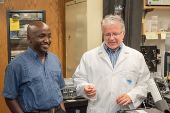
 The clinical report, led by David O. Otuya, PhD is titled, “Passively scanned, single-fiber optical coherence tomography probes for gastrointestinal devices.” The report, published in Lasers in Surgery and Medicine (LSM), the official journal of the American Society for Laser Medicine and Surgery, Inc. (ASLMS), was selected as the July 2022 Editor’s Choice.
“Using methods introduced here, initial imaging identifies an area of interest, providing the location for medical devices to be placed in the gastrointestinal tract,” said Otuya. “Because the medical device enters the same location in which the imaging tool was previously located, this can be considered completing a medical procedure without visual guidance, as is required with the current processes involving endoscopy. This would make procedures cheaper, more convenient, tolerable by patients, and attainable in low resource medical settings.”
David Otuya is a research fellow with the Tearney Laboratory at Massachusetts General Hospital (MGH). His research interest covers topics on biomedical imaging, biopsy and sensing in luminal organs such as the gastrointestinal tract and the airway. He obtained his PhD from Tohoku University, Sendai, Japan, focused on high speed spectrally efficient communication technology in the spring of 2016. Since that time, he has worked with the Tearney lab developing minimally invasive, low-cost devices focused on medical diagnostics tools for low- and middle-income countries (LMICs).
Editor’s Choice is an exclusive article published in LSM, the official journal of the ASLMS. View the complete manuscript.
The American Society for Laser Medicine and Surgery, Inc. (ASLMS) is the largest multidisciplinary professional organization dedicated to the development and application of lasers and related technology for health care applications. ASLMS promotes excellence in patient care by advancing biomedical application of lasers and other related technologies worldwide. ASLMS membership includes physicians, surgeons, nurses, and allied health professionals representing multiple specialties, physicists involved in product development, biomedical engineers, biologists, industry representatives and manufacturers. For more information, visit aslms.org.