Study establishes optical coherence tomography (OCT) as a promising tool for investigating the location and beat frequency of fallopian tube cilia, laying the foundation for future advancements in reproductive physiology.
Wausau, WI (September 29, 2025) – Cilia are tiny, hair-like structures that help transport eggs, embryos, and sperm in the human fallopian tubes, and their impaired movement has been linked to endometriosis, pelvic inflammatory disease, and infertility. Researchers at the University of Arizona and Baylor College of Medicine have demonstrated, for the first time, that optical coherence tomography (OCT) can be used to generate depth-resolved maps and measure the rhythmic beating of human fallopian tube cilia.
The study, led by Dilara J. Long, is titled, “Optical Coherence Tomography Enables the Depth-Resolved Measurement of Cilia Beat Frequency in Ex Vivo Human Fallopian Tubes.” The preclinical study, published in Lasers in Surgery and Medicine (LSM), the official journal of the American Society for Laser Medicine and Surgery, Inc. (ASLMS), was selected as the October 2025 Editor’s Choice.
In this study, fallopian tube tissue was collected from five patients undergoing hysterectomy, and samples were opened to expose the inner surface. OCT imaging was used to create depth-resolved maps of the location and dynamics of cilia in ex vivo human fallopian tubes. The collective movement of cilia was appreciated as a “shimmering” pattern on OCT images. Image processing converted this pattern into measures of beat frequency of groups of cilia.
“Despite the important role of cilia in reproduction, little is known about how they function in the human fallopian tubes,” said Long. “We show that optical coherence tomography (OCT) imaging can reveal the location and beat frequency of surface and hidden fallopian tube cilia, potentially advancing understanding, diagnosis, and management of reproductive disorders.”
These findings establish OCT as a promising tool for studying reproductive physiology and lay the foundation for future development of minimally invasive OCT endoscopes that could one day allow clinicians to directly evaluate cilia function in patients, potentially transforming the diagnosis and management of infertility and related reproductive disorders.
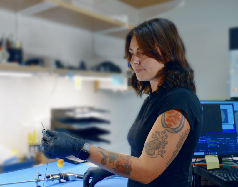
Dilara J. Long is an MD-PhD student at the University of Arizona College of Medicine - Tucson and a PhD candidate in Biomedical Engineering. An aspiring physician-scientist, her research focuses on developing minimally invasive strategies to improve diagnosis and advance understanding of diseases affecting women, including ovarian cancer, endometriosis, and infertility.
Editor’s Choice is an exclusive article published in LSM, the official journal of ASLMS.
View the complete manuscript.