Study introduces a fast, low-cost method that stains heat-damaged skin areas black, allowing thermal injury to be seen directly, rather than inferred from missing tissue signals.
Wausau, WI (March 19, 2026) – A novel staining protocol utilizing standard equipment shows promise as a histological tool in energy-based device (EBD) and burn research, where accurate visualization of collagen damage is essential. According to a recent study, conventional elastic-fiber stain (Verhoeff–van Gieson; VVG) contains a previously underappreciated readout for energy-based thermal injury that renders thermally denatured dermal collagen as a high-contrast black signal while elastic fibers remain distinctly stained within the same section.
-with-heather-m-downs-(left).jpg?sfvrsn=78edad3a_1)
The study, led by Ga Ram Ahn, MD, PhD, is titled, “An Optimized Staining Method for Visualization of Thermally Denatured Dermal Collagen.” The clinical report, published in Lasers in Surgery and Medicine (LSM), the official journal of the American Society for Laser Medicine and Surgery, Inc. (ASLMS), was selected as the March 2026 Editor’s Choice.
“Most histology methods show thermal injury only as a 'missing signal' by staining intact collagen or viable cells,” Ahn said. “We developed a rapid stain that directly highlights thermally denatured collagen plus elastic fiber with dramatic contrast, validated by thermal-imaging dose maps, using common reagents in ~40 minutes on frozen or paraffin sections.”
By deconstructing the VVG workflow step-by-step, the authors identified the ferric-chloride differentiation step as the major source of operator-dependent variability and reduced contrast for the denatured-collagen signal. Omitting this step and optimizing a brief 1% acid-alcohol rinse produced a streamlined protocol, termed Ahn-van Gieson stain (AVG), that preserves elastic-fiber staining while consistently enhancing direct visualization of thermally denatured collagen using standard reagents.
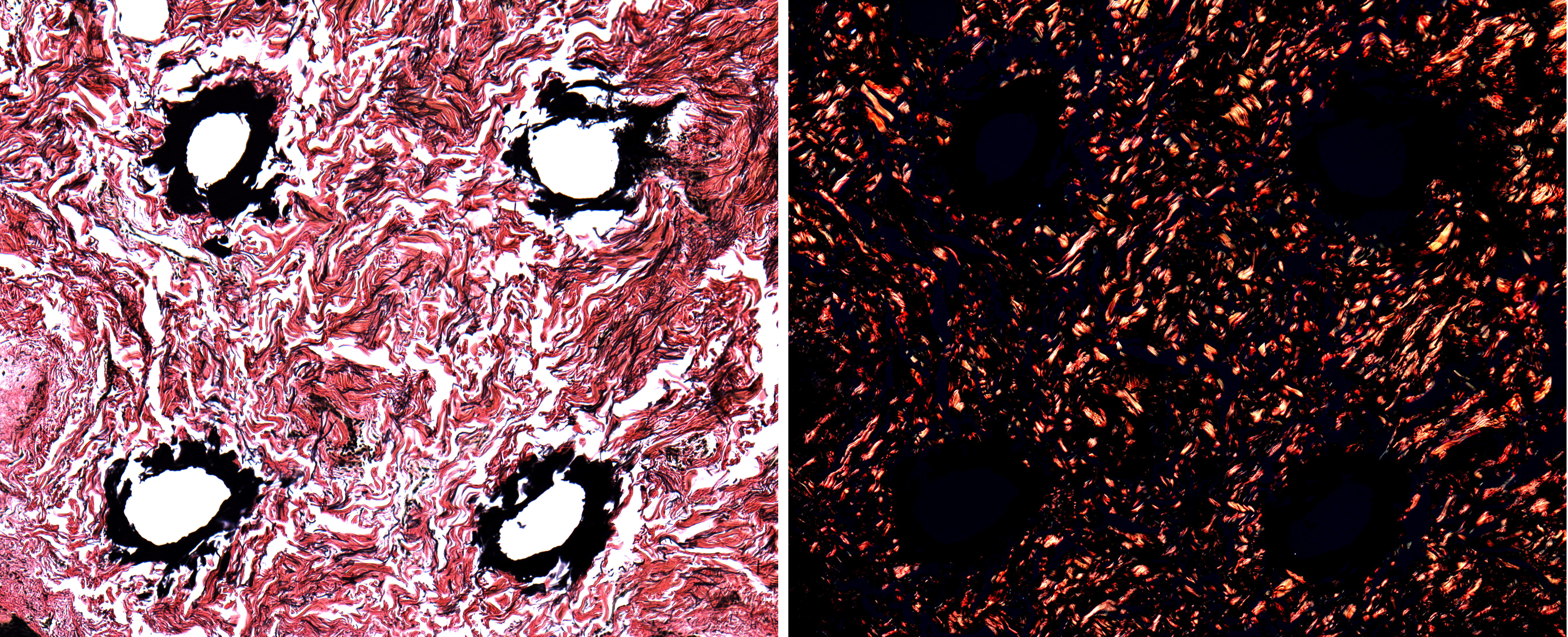
Left: Horizontal section of ex vivo human dermis after CO₂ fractional laser exposure stained with Ahn–van Gieson (AVG), showing discrete black-stained thermal micro-injury zones within the dermal collagen matrix. Right: Corresponding cross-polarized light image demonstrating loss of birefringence at the same micro-injury locations, consistent with collagen denaturation.
Across frozen sections and paraffin blocks, AVG delineated thermal injury zones more clearly and reproducibly than hematoxylin and eosin (H&E), Masson’s trichrome, and nitroblue tetrazolium chloride (NBTC), while remaining compatible with birefringence imaging. The stained regions spatially matched peak-temperature and Arrhenius-integral maps derived from thermal-camera temperature histories, supporting specificity for thermally denatured collagen.
Ga Ram Ahn, MD, PhD, is a dermatologist and instructor in Dermatology at Harvard Medical School and Massachusetts General Hospital (Cutaneous Biology Research Center). His research focuses on dermatologic energy-based device innovation.
Editor’s Choice is an exclusive article published in LSM, the official journal of ASLMS. View the complete manuscript.